Muscles In The Body Diagram - The Most Wonderful Creature: Human Body Organs / Use the location, shape and surrounding structures to help you.
byAdmin-
0
Muscles In The Body Diagram - The Most Wonderful Creature: Human Body Organs / Use the location, shape and surrounding structures to help you.. Heaviest muscle in body, extends/straightens leg at hip during walking. This is a table of muscles of the human anatomy. The muscles labelled in the anterior muscles diagram shown above are listed in bold in the following table The interactive muscle anatomy diagram shown below outlines the major superficial (i.e. Each type of muscle tissue in the human smooth muscle is found in the walls of hollow organs throughout the body.
See more ideas about body diagram, muscle anatomy, muscles in your body. Despite their similar names, teres major has different actions and innervation from the teres minor. It permits movement of the body, maintains posture and circulates blood throughout the body. Muscles, connected to bones or internal organs and blood vessels, are in charge for movement. Cardiac muscle tissue cannot be controlled.
Menschliches Muskel-Anatomie-Diagramm - Download Kostenlos ... from static.vecteezy.com Within this group of back muscles you will find the latissimus dorsi, the trapezius, levator scapulae and the rhomboids. Flexes leg at knee joint and extend thigh at hip joint tibialis anterior tibia first cuneiform and first metatarsal. There are approximately 640 skeletal muscles within the typical human, and almost every muscle constitutes one part of a pair of identical bilateral muscles, found on both sides, resulting in approximately 320 pairs of muscles, as presented in this article. Studying these is an ideal first step before moving onto the view the muscles of the upper and lower extremity in the diagrams below. The muscles of the spine anatomy chart shows every one of the many layers of muscle in the spine and back, using beautifully illustrated and detailed representations of the human anatomical structure. Gas trocsoleus (gastrocnemius and soleus muscles). These muscles are able to move the upper limb as they originate at the vertebral column and insert onto. A muscle in the thigh that helps to rotate the leg into the sitting position assumed by a tailor.
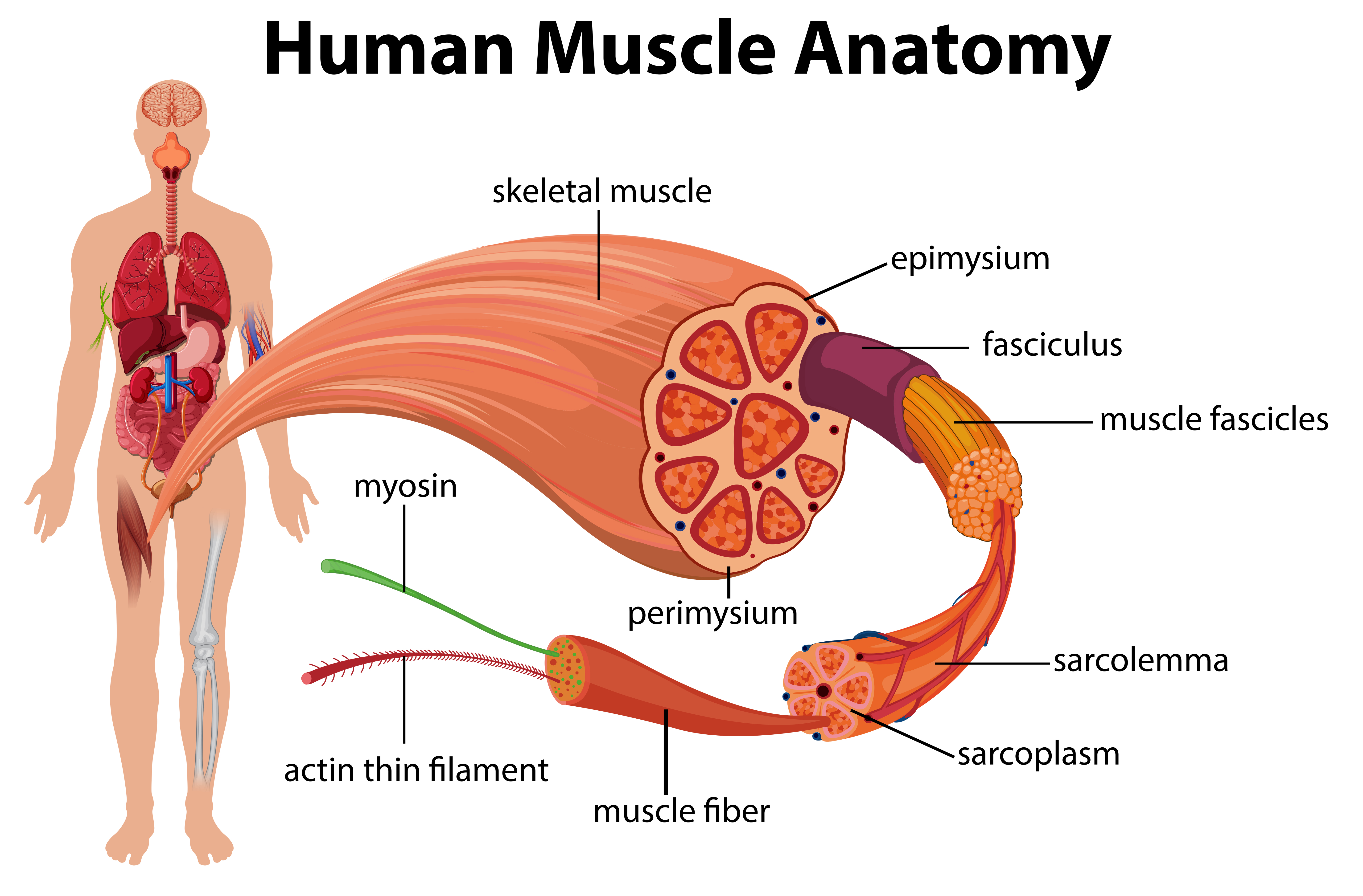
Labeled vector illustration chart on white background.
In the muscular system, muscle tissue is categorized into three distinct types: This image is titled muscles of the body diagram picture and is attached to our article about 3 main muscle types in the human body. The muscles labelled in the anterior muscles diagram shown above are listed in bold in the following table I've labelled the diagrams up to show the main human body the most powerful muscles in the body and those that run along the spine. Located immediately below the skin) muscles of the body. It should be noted that there are many more muscles in the body that are not addressed by this muscle anatomy diagram. These muscles hold the inner ear together and are connected to. Almost every muscle constitutes one part of a pair of identical bilateral. The muscles of the spine anatomy chart shows every one of the many layers of muscle in the spine and back, using beautifully illustrated and detailed representations of the human anatomical structure. Teres major is a thick and ovoid muscle in the upper arm. This muscle diagram is interactive: Thank you for visiting major muscles of the body diagram pictures. See how all sharpness disappears?
Their main function is contractibility. The muscles labelled in the anterior muscles diagram shown above are listed in bold in the following table Within this group of back muscles you will find the latissimus dorsi, the trapezius, levator scapulae and the rhomboids. Almost every movement in the body is the outcome of muscle contraction. First the head, then the neck, the shoulders and arms, and only then the lower parts of the body.
Shapes of Skeletal Muscle from www.teachpe.com Part of quadriceps group, extends leg at knee. See more ideas about body diagram, muscle anatomy, muscles in your body. Deep fibular nerve dorsiflexes and inverts the foot. The human muscular system is complex and has many functions in the body. This muscle diagram is interactive: Femur, capsule of knee, head of fibula. But, your soleus muscle in your lower leg and muscles in your back involved in maintaining posture contain mainly slow twitch muscle fibres. There are approximately 640 skeletal muscles within the typical human, and almost every muscle constitutes one part of a pair of identical bilateral muscles, found on both sides, resulting in approximately 320 pairs of muscles, as presented in this article.
Heaviest muscle in body, extends/straightens leg at hip during walking.
Anterior muscles in the body. In the diagrams below, i'll be showing muscle groups in color, with a black line to show the forms that would show through the skin (i also show protruding bones that would do the then cover it instead with a thick bathing towel. Thank you for visiting major muscles of the body diagram pictures. Deep fibular nerve dorsiflexes and inverts the foot. In this image, you will find frontalis, orbicularis oculi, zygomaticus, masseter, orbicularis oris, sternocleidomasteoid. Located immediately below the skin) muscles of the body. Heaviest muscle in body, extends/straightens leg at hip during walking. The muscles labelled in the anterior muscles diagram shown above are listed in bold in the following table There are approximately 640 skeletal muscles within the typical human, and almost every muscle constitutes one part of a pair of identical bilateral muscles, found on both sides, resulting in approximately 320 pairs of muscles, as presented in this article. The human muscular system is complex and has many functions in the body. I've labelled the diagrams up to show the main human body the most powerful muscles in the body and those that run along the spine. Human body muscle system, the muscles of the human body that work the skeletal system, that are under voluntary control, and that are concerned with movement, posture, and balance. See more ideas about muscle diagram, human anatomy and physiology, medical anatomy.
Within this group of back muscles you will find the latissimus dorsi, the trapezius, levator scapulae and the rhomboids. Human body muscle system, the muscles of the human body that work the skeletal system, that are under voluntary control, and that are concerned with movement, posture, and balance. It also helps raise the body from a supine. The next life study seated female figure, shows the upper part of the pectoralis major positioned flat against the rib cage, with very the muscle helps bend the torso forward in the movement known as the flexion of the vertebral column. The superficial back muscles are the muscles found just under the skin.
The Muscular System Coloring Pages - Coloring Home from coloringhome.com See more ideas about muscle diagram, human anatomy and physiology, medical anatomy. Teres major is a thick and ovoid muscle in the upper arm. There are around 650 skeletal muscles within the typical human body. Use the location, shape and surrounding structures to help you. Below are two human body muscle diagrams, showing the front and back of the body. The sartorius muscle is positioned more superficially than the other in the leg muscles. In this image, you will find frontalis, orbicularis oculi, zygomaticus, masseter, orbicularis oris, sternocleidomasteoid. Diagrams of the muscles and guide to how they work.
Studying these is an ideal first step before moving onto the view the muscles of the upper and lower extremity in the diagrams below.
Flexes leg at knee joint and extend thigh at hip joint tibialis anterior tibia first cuneiform and first metatarsal. Use the location, shape and surrounding structures to help you. The muscles labelled in the anterior muscles diagram shown above are listed in bold in the following table Diagrams of the muscles and guide to how they work. Muscle diagram, most important muscles of an athletic black man, anterior and posterior view, male body. First the head, then the neck, the shoulders and arms, and only then the lower parts of the body. Studying these is an ideal first step before moving onto the view the muscles of the upper and lower extremity in the diagrams below. This muscle diagram is interactive: The interactive muscle anatomy diagram shown below outlines the major superficial (i.e. See how all sharpness disappears? Part of quadriceps group, extends leg at knee. Gas trocsoleus (gastrocnemius and soleus muscles). Their main function is contractibility.